Research
Since the mid 1970s I have been active along the international research front studying scavenger cells, their receptors, and the metabolic fate of the material that these cells clear from the circulation with remarkable speed and capacity. During my PhD work at Uppsala University in the early 1980s I developed a novel method for mass isolation and culture of the two major types of rat liver scavenger cells, the highly specialized Liver Sinusoidal Endothelial Cell (LSEC) and the liver macrophage, or Kupffer Cells (KC). Together these two cell types constitute the most powerful machinery in our body contributing importantly to keep our blood clean (Wikipedia article on the Reticuloendothelial system).
In addition to revealing the importance of both LSECs and KCs as instrumental in the clearance of small and large blood borne compounds in rat, mouse, and other mammals, I contributed to establish that animal species in all vertebrate classes deal with blood clearance in much the same way as mammalian animals, namely by uptake in specialized scavenger endothelial cells and macrophages. However, whereas the liver is the most important site of blood clearance of land based vertebrates, blood clearance in phylogenetically older vertebrates, including bony fishes, cartilagenous fishes, and jawless vertebrates (hagfish and lamprey) is chiefly performed by specialized scavenger endothelial cells and macrophages localized in organs other than liver.
Animations
Dynamic visualisations of the fate of a compound (green) following intravenous injection (Courtesy from D’Liver AS Tromsø, Norway).
Animation 1: Following intravenous injection, the compound travels rapidly with the blood stream. When it reaches the liver, it binds to receptors expressed on the the liver scavenger cells. The yellow “explosions” depict rapid metabolism of the compound following uptake in the cells.
Animation 2: High magnification showing the inside of a liver sinsoid, exposing the surface of liver scavenger cells, with receptors binding the blood borne compond (green). Shown in this animation is also the internalization of the bound compounds, and the return of empty receptors to the cell surface, enabling new rounds of compound binding and internalization. Metabolism of internalized compound is illustrated by yellow explosions.
Infographic
Below is a representation of liver structure shown at increasing magnifications. (Courtesy from D’Liver AS Tromsø, Norway.)
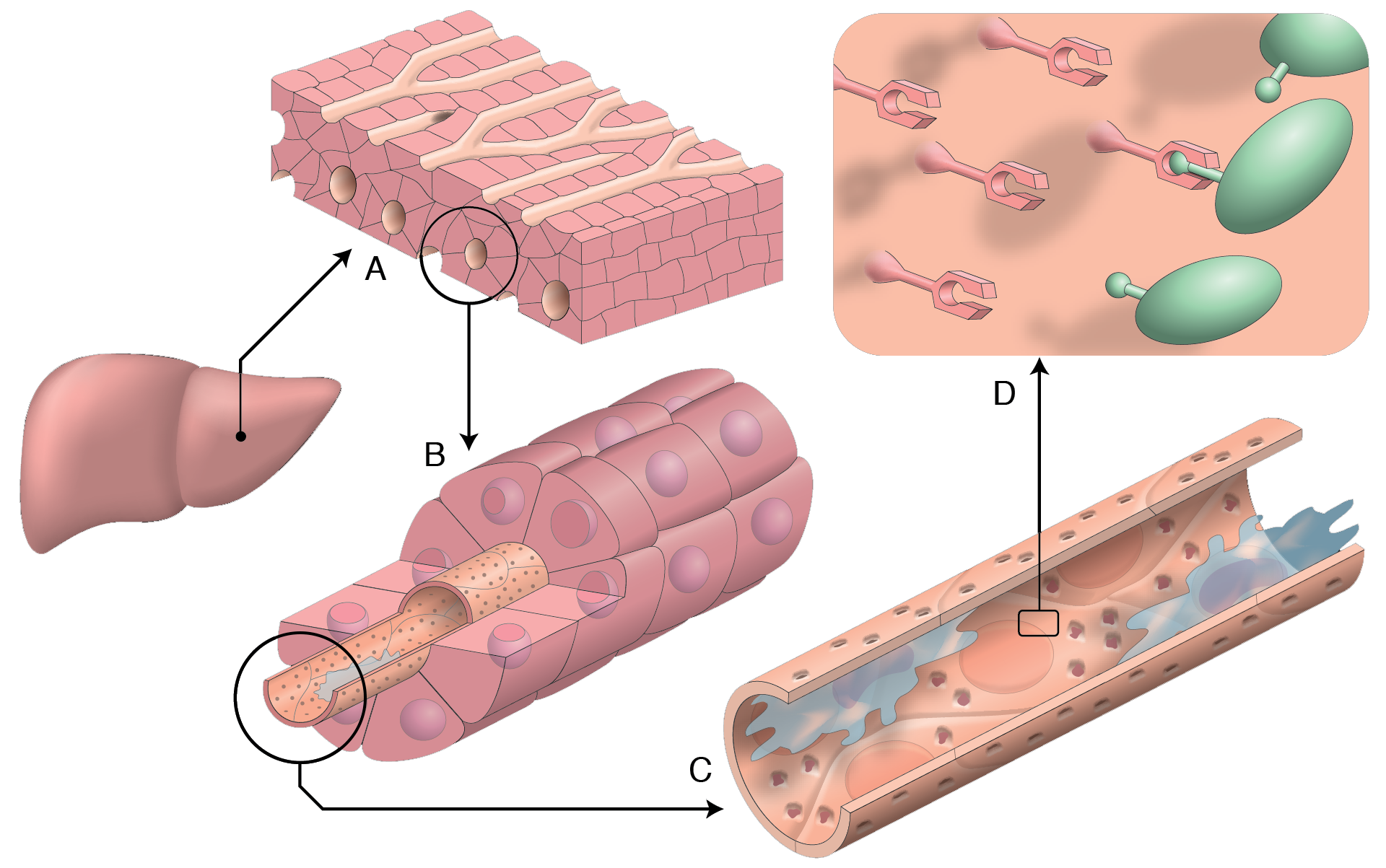
A: The large parenchymal cells (a.k.a. hepatocytes) are depicted as brick like structures surrounding capillaries (a.k.a. liver sinusoids).
B: At higher magnification the cell nuclei of the hepatocytes can be seen. The lining of a sinusoid is detailed.
C: At even higher magnification a sinusoid is shown, with both types of scavenger cells shown (the liver sinusoidal endothelial cells – LSECs) can be seen to make up the lining of the sinusoid, whereas liver macrophages (a.k.a. Kupffer cells – KCs), colored blue, are located on the LSEC lining. At this high magnification the LSECs can be seen to contain nano sized pores (a.k.a. open fenestrae, about 100 nm in diameter).
D: At this submicroscopic magnification clearance receptors on the LSEC surface are shown. One of the receptors has bound a blood borne compound (green) in order to clear it from the circulation (also clearly shown above, in Animation 2). The next event (not shown here) is rapid receptor internalization into the LSEC cell body, bringing the bound compound to the interior of the LSEC to be metabolized.